


Jun 10, 2026 · Press Release
Simplify discovery,
collaborate anywhere.
One tool to manage, explore, annotate, and collaborate on your large microscopy data.
Explore and Collaborate on your Microscopy Datasets from anywhere. Simplified discovery.
Trusted by leading institutions worldwide


All the performance. None of the HURDLES.
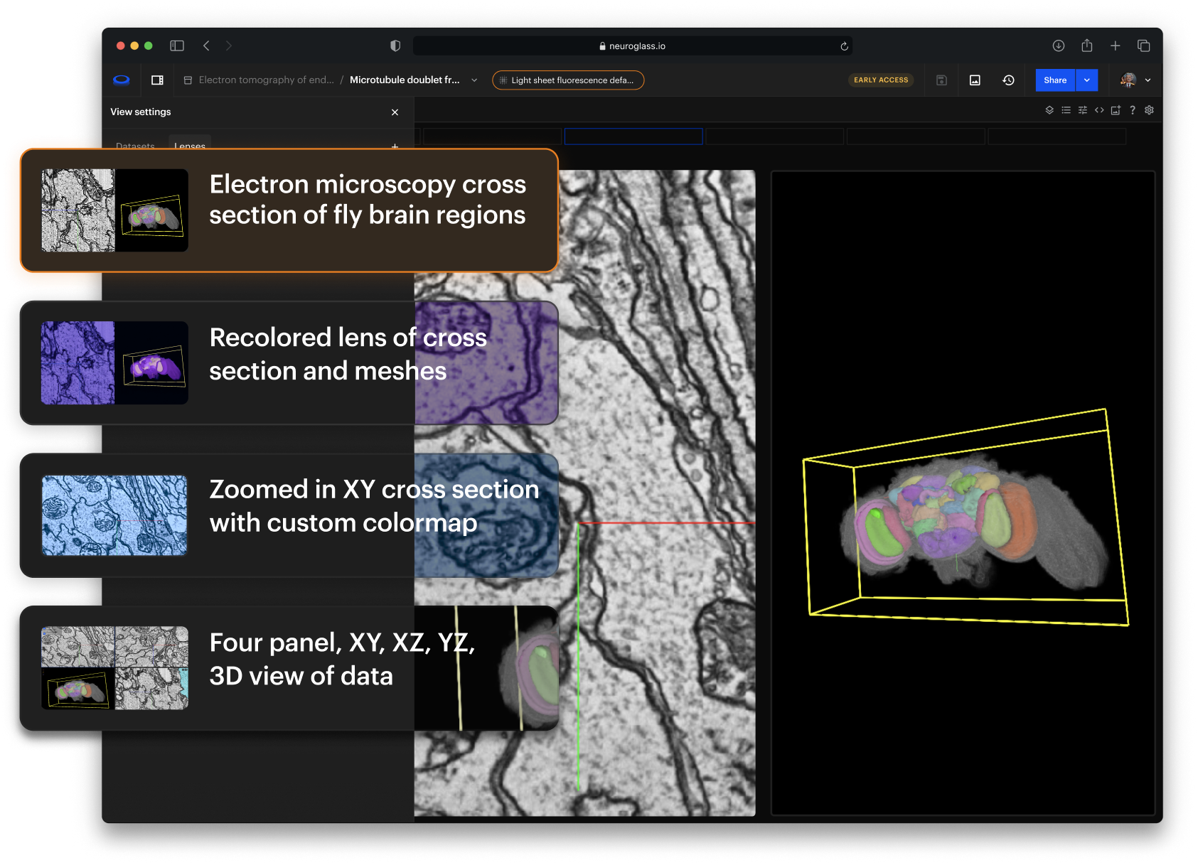
A novel Neuroglancer experience


Attributions: Zheng et al., Cell 2018; Buhmann et al., Nature Methods 2021; Heinrich et al., arXiv 2018
The fastest visualization engine for microscopy data
All the tools and performances of Neuroglancer with an improved user interface and experience.

.gif)
Powerful history
Keep track of everything you do, restore or go back to any previous state. It’s called reproducible science.

Figures. Capture key visualizations from your study
Highlight critical findings and share them with colleagues. Edit and refine whenever you want.

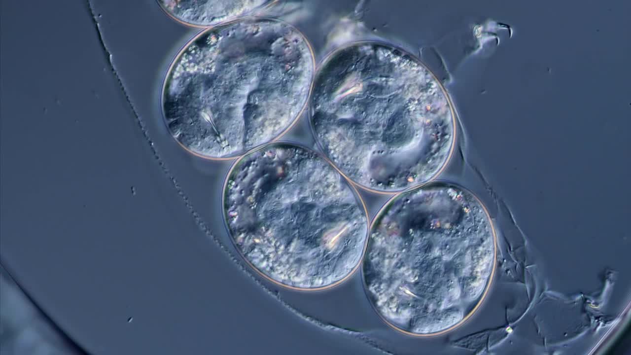
Figure 3 Segmentation of Adult Drosophila Melanogaster
Glances. Reproducible results

Capture a specific moment as you work on your study, preserving key insights for easy reference. Share a reproducible view of your data, enabling collaboration and research continuity.

Designed for your large scale volumetric 3D dataset

Connectomics

Cryo-EM/ET

Expansion Microscopy

High-Res Cellular Imaging


Developmental Imaging

Molecular Imaging
Electron Microscopy
Fluorescence Microscopy
Attributions


Clarity does NOt stop at the image.
Keep track of your data and find what you need

Studies
Organize and visualize your experiments

Visualization engine
All the power of Neuroglancer with an improved user interface and usability experience.
Powerful history
Keep track of everything you do, go back to any previous state. It’s called reproducible science.
Glances
Crystallize any moment and share it seamlessly with other researchers.
Figures
Move, refine, snap. Store and organize the figures for your publications. And bring them back to life with a single click so you can tweak them and refine them.
Glances
Share and publish reproducible results
History
Decide whether NeuroGlass should automatically remember all your activities or if you prefer to manually save specific moments of your study.
Memory
NeuroGlass will remember all the settings and visualisation properties of your navigation in a Glance, so you can go back to it or share it with your collaborators
Share
When you share a Glance, your colleagues will see the exact view that you crafted for them
Publish
Does everyone agree that this Glance is worth sharing with the scientific community? Turn it into a Figure, download it, and include it in your next publication.

Figures
Capture key visualizations from your study
Create
Find the combination of visualisation settings that looks best for you, and create a high resolution snapshot ready for publication.
Edit and re-create
Do you need to make a small change? Every Figure is linked to a Glance, so you can go back to it, edit it and re-create a new version.
Bulk-edits
Coming soon.
Download
Once you are happy with your work, export your high resolution Figure.

Data Collections
Coming soon
Import and organize your datasets

Data schema flexibility
Import the metadata that your community is familiar with. You can add or remove fields to existing data schemas, or create your own!
Easy to use
NeuroGlass will generate a template for you to populate with your data, and re-upload.
Bulk-connect your experiments
All the experiments imported are connected together and managed for you. No more copy and paste.
Speed and scalability
Simplify bulk edits across studies, glances, and figures by applying visualisation settings to all related studies.
Attributions: Zheng et al., Cell 2018; Buhmann et al., Nature Methods 2021; Heinrich et al., arXiv 2018
Metadata
Link your metadata with native OME support or create custom metadata that fits your experiment.

Tagging
Group your Studies, Figures, and Glances with tags for quick, easy access whenever you need them.

Extensive search
Easily find your annotations, layers, Studies and more. NeuroGlass search through your whole database to find what you need.

Put together datasets in the same Collection and switch lenses to explore different perspectives. Keep everything ready in your NeuroGlass dashboard.
NeuroGlass for oRGANIZATIONs
Make it easy for your team to collaborate and publish
NeuroGlass works at scale, removing technical barriers from scientific Studies so your team can focus on the real challenges.

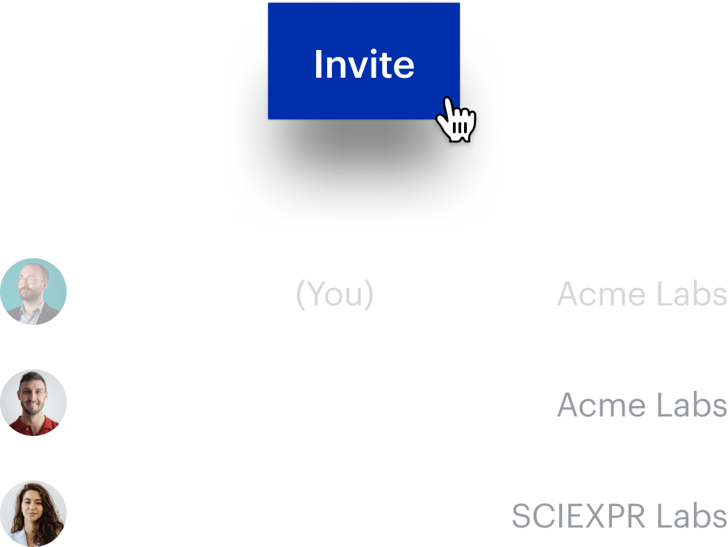
Easily share data within your institution, at scale
Remove the friction, let your team collaborate on annotations the easy way
Make your institution’s data available to the scientific community, as soon as you are ready
Customize NeuroGlass with the logo and colors of your organization
Gather usage statistics for your published Studies
Use NeuroGlass from our own secure deployment or on premise
Use your own fork of Neuroglancer, if needed


Work together. Discover more.
Collaboration should not be hard
Share everything you need with collaborators: simultaneously work on data, looks, annotations, and more
Short URLs
Your Studies can be shared easily with short and manageable links.
Easily share
You can share any content with your colleagues, whether they are NeuroGlass users or not.
Live presence
Collaborate on the same Study in real time. Share ideas and get inspired.
Ensuring data security
Private S3 bucket integration for secure storage.
Connect and read data directly from private S3 buckets, allowing you to keep them in controlled storage.

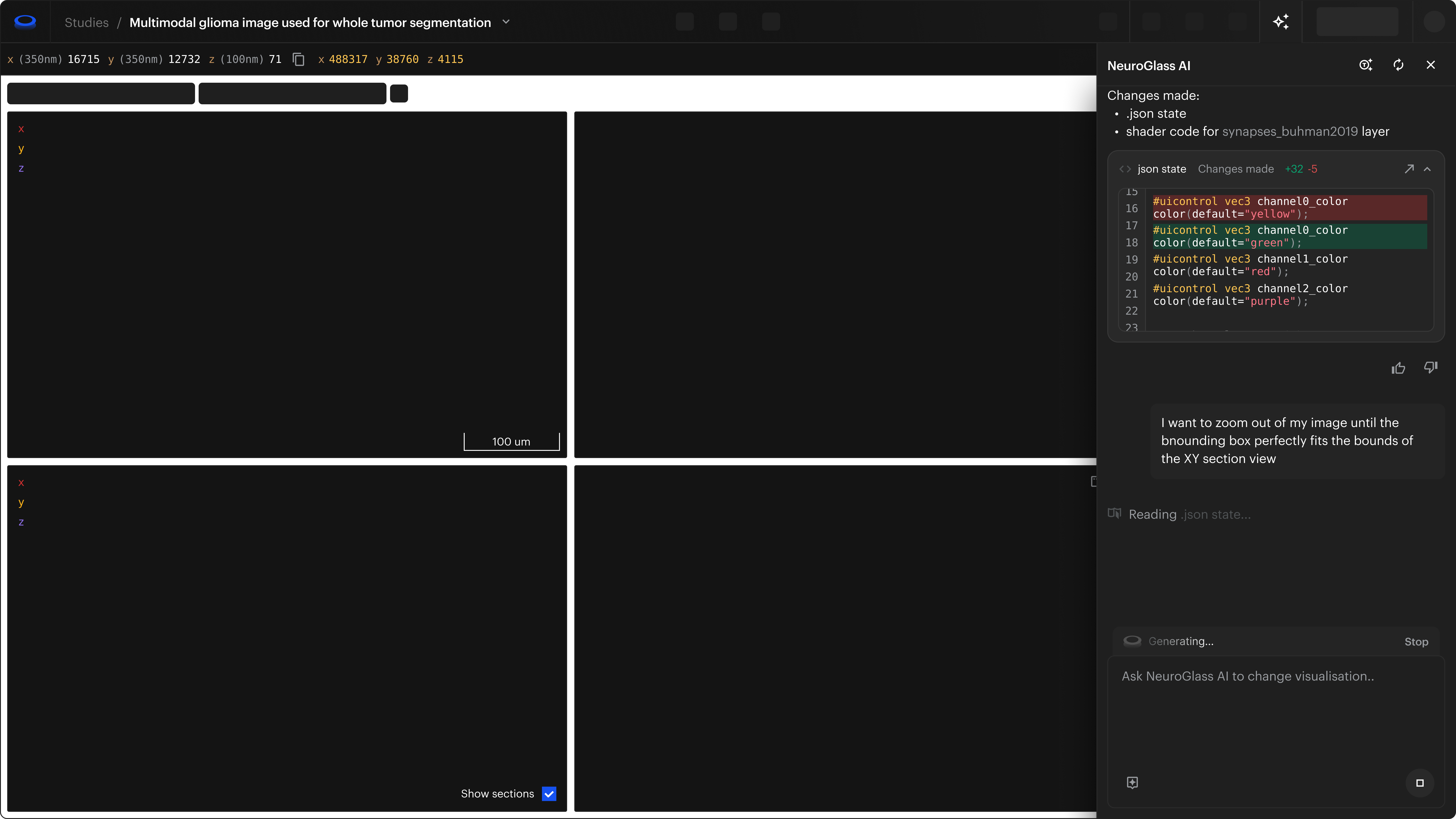
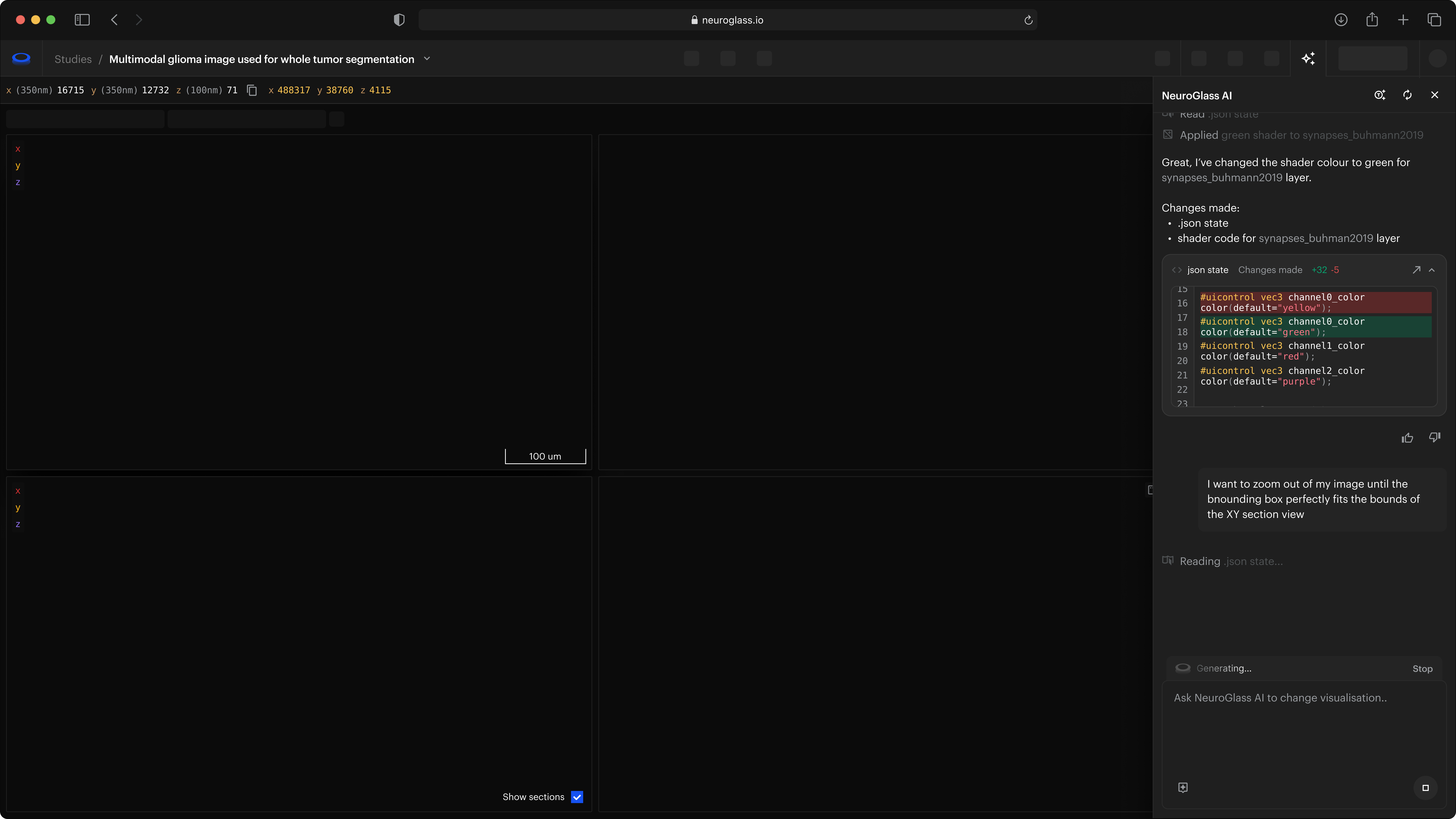
Automate. Integrate. Innovate.
Built for scientists. Ready for developers.
Seamlessly import your data or programmatically create your Studies with our developer-friendly APIs.
Visit API documentation


Your data, protected by design.
Research securely. Collaborate confidently.
We take your data security seriously — that’s why we ensure it never leaves its trusted home.
Secure hosting
Whether deployed in your institution or on MetaCell Cloud, your research data never leaves your servers.
GDPR compliance
Your information is safe with us. Visit the Privacy Terms to know more about how we treat personal information.
Secure Single sign-on (SSO)
Integrate NeuroGlass with your authentication provider for easy access.


What is coming next
We are actively working to improve NeuroGlass. Want to fill in the blocks below?
Let us know what features you'd like to see
Comments
Tasks
Skeleton Tracing
Segmentation
Data conversion
Data preparation

Book your office hours slot
Join for 1-hour live session to see NeuroGlass in action.
We'll walk through core features, real workflows, and the tools researchers use every day.
We'll walk through core features, real workflows, and the tools researchers use every day.
Reserve your spot

Frequently Asked Questions
Can't find an answer? Contact us
What is NeuroGlass?
What data does NeuroGlass support?
How do I import my data into NeuroGlass
When I create a Data Collection, do I need to upload the raw image files?
Is my data secure in NeuroGlass?
Can I collaborate with my team using NeuroGlass?
Can I use NeuroGlass free of charge?
Contributing back to the Neuroglancer community
We built NeuroGlass to accelerate research breakthroughs by simplifying collaboration and enhancing productivity. NeuroGlass wouldn't exist without Neuroglancer and the incredible contributions of its community.
NeuroGlass is committed to giving back—keeping all Neuroglancer improvements open source and freely available for everyone.
NeuroGlass is committed to giving back—keeping all Neuroglancer improvements open source and freely available for everyone.

Step into the future of research
Explore and Collaborate on your Microscopy Datasets from anywhere. Simplified discovery.


Copyright © 2026 MetaCell LLC, LTD. All rights reserved.
Neuroglass.io and Neuroglass.com are owned and operated by MetaCell, LLC. MetaCell.us contains copyrighted and other proprietary information. Copyright on Neuroglass.io and Neuroglass.com (including but not limited to text, photographs, graphics, videos and software) is owned by MetaCell or its partners. No right, title or interest in any content of Neuroglass.io and Neuroglass.com is transferred or licensed to you as a result of your use of the website. Other than for private use, you may not download or copy, store in any medium (including any other website), distribute, transmit, re-transmit, modify, show in public or make commercial or business use of any part of Neuroglass.io and Neuroglass.com without the prior written consent of MetaCell. By accessing Neuroglass.io and Neuroglass.com you agree not to use the content for any illegal or improper purpose. No part of Neuroglass.io and Neuroglass.com may be reproduced or distributed in any material form or medium without the permission of MetaCell.
Neuroglass.io and Neuroglass.com are owned and operated by MetaCell, LLC. MetaCell.us contains copyrighted and other proprietary information. Copyright on Neuroglass.io and Neuroglass.com (including but not limited to text, photographs, graphics, videos and software) is owned by MetaCell or its partners. No right, title or interest in any content of Neuroglass.io and Neuroglass.com is transferred or licensed to you as a result of your use of the website. Other than for private use, you may not download or copy, store in any medium (including any other website), distribute, transmit, re-transmit, modify, show in public or make commercial or business use of any part of Neuroglass.io and Neuroglass.com without the prior written consent of MetaCell. By accessing Neuroglass.io and Neuroglass.com you agree not to use the content for any illegal or improper purpose. No part of Neuroglass.io and Neuroglass.com may be reproduced or distributed in any material form or medium without the permission of MetaCell.
Allen Institute using NeuroGlass to support large-scale brain imaging workflows
Read the Press Release